Review


gst alk protein  (Carna Inc)


Bioz Verified Symbol Carna Inc is a verified supplier
Bioz Manufacturer Symbol Carna Inc manufactures this product  
  • Logo
  • About
  • News
  • Press Release
  • Team
  • Advisors
  • Partners
  • Contact
  • Bioz Stars
  • Bioz vStars
  • 95

    Structured Review

    Carna Inc gst alk protein
    Gst Alk Protein, supplied by Carna Inc, used in various techniques. Bioz Stars score: 95/100, based on 24 PubMed citations. ZERO BIAS - scores, article reviews, protocol conditions and more
    https://www.bioz.com/product/gst+alk+protein/ALK/us10980816-875-5-12
    Average 95 stars, based on 24 article reviews
    gst alk protein - by Bioz Stars, 2026-09
    95/100 stars

    Images

    Related Articles

    Mutagenesis:

    Article Title: Thienopyrimidine derivatives, processes for the preparation thereof and therapeutic uses thereof
    Article Snippet: The protocol is the same as the one for the wild-type form, but the final ATP concentration is 200 microM and the final enzyme concentration is 1 nM.

    Article Title: Thienopyrimidine derivatives, processes for the preparation thereof and therapeutic uses thereof
    Article Snippet: The protocol is the same as the one for the wild-type form, but the final ATP concentration is 200 microM and the final enzyme concentration is 1 nM.

    Article Title: Thienopyrimidine derivatives, processes for the preparation thereof and therapeutic uses thereof
    Article Snippet: The reagents used have the following composition: Enzyme buffer (EB): 50 mM HEPES (pH: 7.0) (Sigma H7523), 100 mM NaCl (Sigma S7653), NaN3 at 0.01% (Sigma S8032), BSA at 0.005% (Sigma A2153), 0.05 mM sodium orthovanadate (Calbiochem 567540).

    Article Title: Thienopyrimidine derivatives, processes for the preparation thereof and therapeutic uses thereof
    Article Snippet: The reagents used have the following composition: Enzyme buffer (EB): 50 mM HEPES (pH: 7.0) (Sigma H7523), 100 mM NaCl (Sigma S7653), NaN3 at 0.01% (Sigma S8032), BSA at 0.005% (Sigma A2153), 0.05 mM sodium orthovanadate (Calbiochem 567540).

    Article Title: Thienopyrimidine derivatives, processes for the preparation thereof and therapeutic uses thereof
    Article Snippet: The reagents used have the following composition: Enzyme buffer (EB): 50 mM HEPES (pH: 7.0) (Sigma H7523), 100 mM NaCl (Sigma S7653), NaN3 at 0.01% (Sigma S8032), BSA at 0.005% (Sigma A2153), 0.05 mM sodium orthovanadate (Calbiochem 567540).

    Article Title: Thienopyrimidine derivatives, processes for the preparation thereof and therapeutic uses thereof
    Article Snippet: The reagents used have the following composition: Enzyme buffer (EB): 50 mM HEPES (pH: 7.0) (Sigma H7523), 100 mM NaCl (Sigma S7653), NaN3 at 0.01% (Sigma S8032), BSA at 0.005% (Sigma A2153).

    Article Title: Thienopyrimidine derivatives, processes for the preparation thereof and therapeutic uses thereof
    Article Snippet: The protocol is the same as the one for the wild-type form, but the final ATP concentration is 200 microM and the final enzyme concentration is 1 nM.

    Article Title: Thienopyrimidine derivatives, processes for the preparation thereof and therapeutic uses thereof
    Article Snippet: The protocol is the same as the one for the wild-type form, but the final ATP concentration is 200 microM and the final enzyme concentration is 1 nM.


    Similar Products

    95
    Carna Inc gst alk protein
    Gst Alk Protein, supplied by Carna Inc, used in various techniques. Bioz Stars score: 95/100, based on 1 PubMed citations. ZERO BIAS - scores, article reviews, protocol conditions and more
    https://www.bioz.com/product/gst+alk+protein/ALK/us10980816-875-5-12
    Average 95 stars, based on 1 article reviews
    gst alk protein - by Bioz Stars, 2026-09
    95/100 stars
      Buy from Supplier

    94
    SignalChem gst alk
    (A) Brain lysates from 4 individual mice of the indicated genotypes were subjected to immunoblotting with phospho-IGF-1R (pIGF-1R Y1135/1136 ) and Gapdh as control to verify equal protein loading. Quantitation of band intensities for pIGF-1R/Gapdh as fold change relative to control WT3 (WT, mouse #3) is plotted. (B) The IGF-1R inhibitor, PPP rescues the multiple axon phenotype in cortical neurons from <t>Alk</t> or Ltk knockout mice. Cortical neurons, isolated from Alk or Ltk knockout mice were treated with varying concentrations of PPP or DMSO as control 4h after plating, fixed and stained for Tuj1. Quantitation of the percent of neurons with multiple, single or no axon-like projections is plotted as the mean ± SD, n = 100 neurons from 2 independent experiments. (C) Analysis of the effect of ectopically applied recombinant IGF-1. Cortical neurons isolated from WT or DKO mice electroporated with a GFP plasmid were treated with PPP (200 nM), DMSO as control and /or IGF-1 (20 or 50 nM), insulin (100 nM) or EGF (20 ng/ml). Cells were fixed at 33h and GFP positive cells were analyzed. Quantitation of the percent of neurons with multiple, single or no axon-like projections is plotted as the mean ± SD, n=100 neurons from 2 (for EGF) or n=200 neurons from 4 (remainder) independent experiments. Data in B and C were analyzed using one-way ANOVA and comparisons to control were performed using Dunnett’s test (**** p<0.0001, *** p<0.001). (D) Expression of Igf-1r mRNA was determined by qPCR in cortical neurons from WT and knockout mice. Data are plotted as mean ± SD of 3 independent experiments. (E-F) IGF-1R associates with LTK and ALK in HEK293T cells. Lysates from HEK 293T cells, transfected with IGF-1R-Flag, LTK-GFP (E) or ALK-GFP (F) were subjected to immunoprecipitation using anti-Flag antibody and co-precipitated LTK (E) or ALK (F) was detected by immunoblotting using anti-GFP antibody. (G) LTK activity is required for interaction with IGF-1R. HEK293T cells were transfected with wild type (WT) or kinase inactive (KR) versions of LTK-Flag. Endogenous IGF-1R was immunoprecipitated with anti-IGF-1R antibody and co-precipitated LTK-Flag was detected by immunoblotting with anti-Flag antibody. (H) LTK/ALK interacts with WT and kinase inactive (KR) variants of IGF-1R. Lysates from HEK293T cells, co-transfected with LTK-WT-HA and WT or KR versions of IGF-1R-Flag were subjected to immunoprecipitation with anti-Flag antibody and co-precipitated LTK was detected by immunoblotting with anti-HA antibody. (I) <t>GST-LTK</t> immobilized to GSH-Sepharose beads was incubated with recombinant His-IGF-1R. Pull down and totals were subjected to immunoblotting using indicated antibodies. (J) Purified recombinant GST-LTK interacts with endogenous Igf-1r. Lysates from WT embryonic brains were incubated with GST-LTK or GST alone, immobilized to GSH-Sepharose beads. Bound and total proteins were subjected to immunoblotting with anti-IGF-1R antibody. (E-J) A representative result from 3 independent experiments is shown.
    Gst Alk, supplied by SignalChem, used in various techniques. Bioz Stars score: 94/100, based on 1 PubMed citations. ZERO BIAS - scores, article reviews, protocol conditions and more
    https://www.bioz.com/product/gst+alk+protein/GST+Protein/bio_rxiv__2023__01__29__526107-275-12-13
    Average 94 stars, based on 1 article reviews
    gst alk - by Bioz Stars, 2026-09
    94/100 stars
      Buy from Supplier

    Image Search Results


    (A) Brain lysates from 4 individual mice of the indicated genotypes were subjected to immunoblotting with phospho-IGF-1R (pIGF-1R Y1135/1136 ) and Gapdh as control to verify equal protein loading. Quantitation of band intensities for pIGF-1R/Gapdh as fold change relative to control WT3 (WT, mouse #3) is plotted. (B) The IGF-1R inhibitor, PPP rescues the multiple axon phenotype in cortical neurons from Alk or Ltk knockout mice. Cortical neurons, isolated from Alk or Ltk knockout mice were treated with varying concentrations of PPP or DMSO as control 4h after plating, fixed and stained for Tuj1. Quantitation of the percent of neurons with multiple, single or no axon-like projections is plotted as the mean ± SD, n = 100 neurons from 2 independent experiments. (C) Analysis of the effect of ectopically applied recombinant IGF-1. Cortical neurons isolated from WT or DKO mice electroporated with a GFP plasmid were treated with PPP (200 nM), DMSO as control and /or IGF-1 (20 or 50 nM), insulin (100 nM) or EGF (20 ng/ml). Cells were fixed at 33h and GFP positive cells were analyzed. Quantitation of the percent of neurons with multiple, single or no axon-like projections is plotted as the mean ± SD, n=100 neurons from 2 (for EGF) or n=200 neurons from 4 (remainder) independent experiments. Data in B and C were analyzed using one-way ANOVA and comparisons to control were performed using Dunnett’s test (**** p<0.0001, *** p<0.001). (D) Expression of Igf-1r mRNA was determined by qPCR in cortical neurons from WT and knockout mice. Data are plotted as mean ± SD of 3 independent experiments. (E-F) IGF-1R associates with LTK and ALK in HEK293T cells. Lysates from HEK 293T cells, transfected with IGF-1R-Flag, LTK-GFP (E) or ALK-GFP (F) were subjected to immunoprecipitation using anti-Flag antibody and co-precipitated LTK (E) or ALK (F) was detected by immunoblotting using anti-GFP antibody. (G) LTK activity is required for interaction with IGF-1R. HEK293T cells were transfected with wild type (WT) or kinase inactive (KR) versions of LTK-Flag. Endogenous IGF-1R was immunoprecipitated with anti-IGF-1R antibody and co-precipitated LTK-Flag was detected by immunoblotting with anti-Flag antibody. (H) LTK/ALK interacts with WT and kinase inactive (KR) variants of IGF-1R. Lysates from HEK293T cells, co-transfected with LTK-WT-HA and WT or KR versions of IGF-1R-Flag were subjected to immunoprecipitation with anti-Flag antibody and co-precipitated LTK was detected by immunoblotting with anti-HA antibody. (I) GST-LTK immobilized to GSH-Sepharose beads was incubated with recombinant His-IGF-1R. Pull down and totals were subjected to immunoblotting using indicated antibodies. (J) Purified recombinant GST-LTK interacts with endogenous Igf-1r. Lysates from WT embryonic brains were incubated with GST-LTK or GST alone, immobilized to GSH-Sepharose beads. Bound and total proteins were subjected to immunoblotting with anti-IGF-1R antibody. (E-J) A representative result from 3 independent experiments is shown.

    Journal: bioRxiv

    Article Title: LTK and ALK regulate neuronal polarity and cortical migration by modulating IGF1R activity

    doi: 10.1101/2023.01.29.526107

    Figure Lengend Snippet: (A) Brain lysates from 4 individual mice of the indicated genotypes were subjected to immunoblotting with phospho-IGF-1R (pIGF-1R Y1135/1136 ) and Gapdh as control to verify equal protein loading. Quantitation of band intensities for pIGF-1R/Gapdh as fold change relative to control WT3 (WT, mouse #3) is plotted. (B) The IGF-1R inhibitor, PPP rescues the multiple axon phenotype in cortical neurons from Alk or Ltk knockout mice. Cortical neurons, isolated from Alk or Ltk knockout mice were treated with varying concentrations of PPP or DMSO as control 4h after plating, fixed and stained for Tuj1. Quantitation of the percent of neurons with multiple, single or no axon-like projections is plotted as the mean ± SD, n = 100 neurons from 2 independent experiments. (C) Analysis of the effect of ectopically applied recombinant IGF-1. Cortical neurons isolated from WT or DKO mice electroporated with a GFP plasmid were treated with PPP (200 nM), DMSO as control and /or IGF-1 (20 or 50 nM), insulin (100 nM) or EGF (20 ng/ml). Cells were fixed at 33h and GFP positive cells were analyzed. Quantitation of the percent of neurons with multiple, single or no axon-like projections is plotted as the mean ± SD, n=100 neurons from 2 (for EGF) or n=200 neurons from 4 (remainder) independent experiments. Data in B and C were analyzed using one-way ANOVA and comparisons to control were performed using Dunnett’s test (**** p<0.0001, *** p<0.001). (D) Expression of Igf-1r mRNA was determined by qPCR in cortical neurons from WT and knockout mice. Data are plotted as mean ± SD of 3 independent experiments. (E-F) IGF-1R associates with LTK and ALK in HEK293T cells. Lysates from HEK 293T cells, transfected with IGF-1R-Flag, LTK-GFP (E) or ALK-GFP (F) were subjected to immunoprecipitation using anti-Flag antibody and co-precipitated LTK (E) or ALK (F) was detected by immunoblotting using anti-GFP antibody. (G) LTK activity is required for interaction with IGF-1R. HEK293T cells were transfected with wild type (WT) or kinase inactive (KR) versions of LTK-Flag. Endogenous IGF-1R was immunoprecipitated with anti-IGF-1R antibody and co-precipitated LTK-Flag was detected by immunoblotting with anti-Flag antibody. (H) LTK/ALK interacts with WT and kinase inactive (KR) variants of IGF-1R. Lysates from HEK293T cells, co-transfected with LTK-WT-HA and WT or KR versions of IGF-1R-Flag were subjected to immunoprecipitation with anti-Flag antibody and co-precipitated LTK was detected by immunoblotting with anti-HA antibody. (I) GST-LTK immobilized to GSH-Sepharose beads was incubated with recombinant His-IGF-1R. Pull down and totals were subjected to immunoblotting using indicated antibodies. (J) Purified recombinant GST-LTK interacts with endogenous Igf-1r. Lysates from WT embryonic brains were incubated with GST-LTK or GST alone, immobilized to GSH-Sepharose beads. Bound and total proteins were subjected to immunoblotting with anti-IGF-1R antibody. (E-J) A representative result from 3 independent experiments is shown.

    Article Snippet: For binding assays, recombinant GST (SignalChem #G52-30U) and GST-LTK (SignalChem #L11-11G) or GST-ALK (SignalChem #A19-116) were incubated with GSH-Sepharose beads (GE Healthcare) in 400 µl binding buffer (50 mM Tris, pH 7.4, 150 mM NaCl, 1 mM EDTA and 0.5% NP-40) for 1 h at 4°C.

    Techniques: Western Blot, Quantitation Assay, Knock-Out, Isolation, Staining, Recombinant, Plasmid Preparation, Expressing, Transfection, Immunoprecipitation, Activity Assay, Incubation, Purification

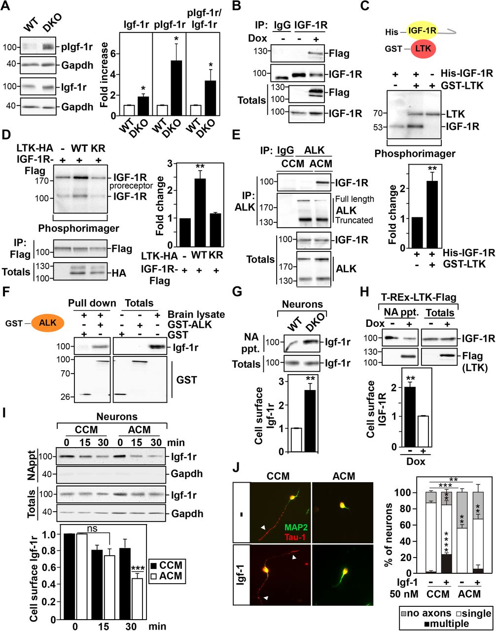
    (A) Representative immunoblot showing the levels of phosphorylated (pIgf-1r Y1135/1136 ), total Igf-1r and Gapdh (as loading control) in lysates of isolated cortical neurons from WT and DKO mice. Bands intensities for pIgf-1r and Igf-1r were normalized to Gapdh and the fold increase relative to WT is plotted as the mean ± SEM of 3 independent experiments. (B) Endogenous IGF-1R was immunoprecipitated from cell lysates of T-REx cells, treated without (-) or with (+) doxycycline (Dox) to induce expression of Flag-tagged LTK and the interaction with LTK-Flag was visualized by immunoblotting. (C) Representative image showing phosphorylation of recombinant His-tagged IGF-1R (kinase domain and C-terminal region) by recombinant GST-LTK (kinase domain) in an in vitro radioactive kinase assay. Phosphorylation of IGF-1R was quantitated as fold change relative to auto-phosphorylation of IGF-1R alone and plotted as mean ± SEM from 7 independent experiments. (D) Representative image showing the phosphorylation of IGF-1R by co-expressed WT or Kinase-deficient (KR) LTK in in vitro radioactive kinase assay. The levels of immunoprecipitated IGF-1R and total LTK-HA were visualized by immunobloting (bottom). Phosphorylation of IGF-1R was quantitated as fold change relative to auto-phosphorylation of IGF-1R alone and plotted as mean ± SEM from 5 independent experiments. (E) Endogenous ALK was immunoprecipitated from lysates of IMR-32 cells, treated with control (CCM) or ALKAL2 (ACM) conditioned media and co-precipitated endogenous IGF-1R was detected by immunoblotting. (F) Homogenates from WT embryonic brains were incubated with GST-ALK or GST alone, immobilized to GSH-Sepharose beads. Bound and total proteins were subjected to immunoblotting with anti-IGF-1R antibody. (G) Cell-surface expression of Igf-1r is increased in DKO cortical neurons. Cell-surface Igf-1r (NA ppt) is normalized to totals and the fold change relative to WT samples is plotted as the mean ± SEM from three independent experiments. (H) LTK decreases cell-surface expression of IGF-1R in T-Rex-LTK stable cells. Cell-surface IGF-1R (NA ppt) normalized to totals as fold change relative to samples expressing LTK (+Dox) is plotted as the mean ± SEM from 3 independent experiments. (I) WT cortical neurons were labeled with biotin and incubated at 37°C for 15 or 30 min in the presence of CCM or ACM conditioned media. Cell-surface Igf-1r (NA ppt) normalized to totals as the fold change relative to controls at time 0 is plotted as the mean ± SEM from 4 independent experiments. (J) WT and DKO cortical neurons were treated with control (CCM) or ALKAL2 (ACM) conditioned medium 4h after plating, and IGF-1 was added 1 hour later. Representative images of cells fixed at 36 h and stained for Tau-1 (axons; red) and MAP2 (dendrites: green) are shown. Arrowheads mark axons. Quantitation of the percent of neurons with multiple, single or no axons is plotted as the mean ± SEM of n = 150 neurons per condition from 3 independent experiments. (A-B, E-G) Representative blots from 3 independent experiments are shown. Statistical significance: ****p<0.0001, ***p<0.001; **p<0.01; *p<0.05 by Student’s t-test.

    Journal: bioRxiv

    Article Title: LTK and ALK regulate neuronal polarity and cortical migration by modulating IGF1R activity

    doi: 10.1101/2023.01.29.526107

    Figure Lengend Snippet: (A) Representative immunoblot showing the levels of phosphorylated (pIgf-1r Y1135/1136 ), total Igf-1r and Gapdh (as loading control) in lysates of isolated cortical neurons from WT and DKO mice. Bands intensities for pIgf-1r and Igf-1r were normalized to Gapdh and the fold increase relative to WT is plotted as the mean ± SEM of 3 independent experiments. (B) Endogenous IGF-1R was immunoprecipitated from cell lysates of T-REx cells, treated without (-) or with (+) doxycycline (Dox) to induce expression of Flag-tagged LTK and the interaction with LTK-Flag was visualized by immunoblotting. (C) Representative image showing phosphorylation of recombinant His-tagged IGF-1R (kinase domain and C-terminal region) by recombinant GST-LTK (kinase domain) in an in vitro radioactive kinase assay. Phosphorylation of IGF-1R was quantitated as fold change relative to auto-phosphorylation of IGF-1R alone and plotted as mean ± SEM from 7 independent experiments. (D) Representative image showing the phosphorylation of IGF-1R by co-expressed WT or Kinase-deficient (KR) LTK in in vitro radioactive kinase assay. The levels of immunoprecipitated IGF-1R and total LTK-HA were visualized by immunobloting (bottom). Phosphorylation of IGF-1R was quantitated as fold change relative to auto-phosphorylation of IGF-1R alone and plotted as mean ± SEM from 5 independent experiments. (E) Endogenous ALK was immunoprecipitated from lysates of IMR-32 cells, treated with control (CCM) or ALKAL2 (ACM) conditioned media and co-precipitated endogenous IGF-1R was detected by immunoblotting. (F) Homogenates from WT embryonic brains were incubated with GST-ALK or GST alone, immobilized to GSH-Sepharose beads. Bound and total proteins were subjected to immunoblotting with anti-IGF-1R antibody. (G) Cell-surface expression of Igf-1r is increased in DKO cortical neurons. Cell-surface Igf-1r (NA ppt) is normalized to totals and the fold change relative to WT samples is plotted as the mean ± SEM from three independent experiments. (H) LTK decreases cell-surface expression of IGF-1R in T-Rex-LTK stable cells. Cell-surface IGF-1R (NA ppt) normalized to totals as fold change relative to samples expressing LTK (+Dox) is plotted as the mean ± SEM from 3 independent experiments. (I) WT cortical neurons were labeled with biotin and incubated at 37°C for 15 or 30 min in the presence of CCM or ACM conditioned media. Cell-surface Igf-1r (NA ppt) normalized to totals as the fold change relative to controls at time 0 is plotted as the mean ± SEM from 4 independent experiments. (J) WT and DKO cortical neurons were treated with control (CCM) or ALKAL2 (ACM) conditioned medium 4h after plating, and IGF-1 was added 1 hour later. Representative images of cells fixed at 36 h and stained for Tau-1 (axons; red) and MAP2 (dendrites: green) are shown. Arrowheads mark axons. Quantitation of the percent of neurons with multiple, single or no axons is plotted as the mean ± SEM of n = 150 neurons per condition from 3 independent experiments. (A-B, E-G) Representative blots from 3 independent experiments are shown. Statistical significance: ****p<0.0001, ***p<0.001; **p<0.01; *p<0.05 by Student’s t-test.

    Article Snippet: For binding assays, recombinant GST (SignalChem #G52-30U) and GST-LTK (SignalChem #L11-11G) or GST-ALK (SignalChem #A19-116) were incubated with GSH-Sepharose beads (GE Healthcare) in 400 µl binding buffer (50 mM Tris, pH 7.4, 150 mM NaCl, 1 mM EDTA and 0.5% NP-40) for 1 h at 4°C.

    Techniques: Western Blot, Isolation, Immunoprecipitation, Expressing, Recombinant, In Vitro, Kinase Assay, Incubation, Labeling, Staining, Quantitation Assay